
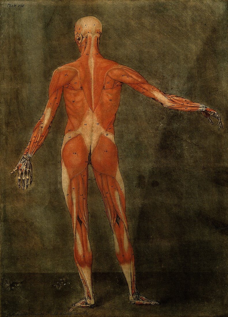
Look at the picture above. It was printed in 1773, copied from the work of the Dutch anatomist Bernhard Siegfried Albinus. Every muscle of the back is there, down to the last fibre. The buttocks are drawn as two great rounded masses, bigger than almost anything else on the body.
By then, anatomists had cut open bodies for more than two hundred years. They knew the shape of the glutes, where they start on the pelvis and where they end on the thigh. What they did not know was what the muscle does in a living person, and when. A dead body shows you the shape. It does not show you the job.
This chapter is about three men who tried to answer that question in the 1800s. One used logic. One used electricity. One used a sick child's walk. Between them, they set up a debate that took another hundred years to settle.
Part 1 · The idea
"The only animal with true buttocks"
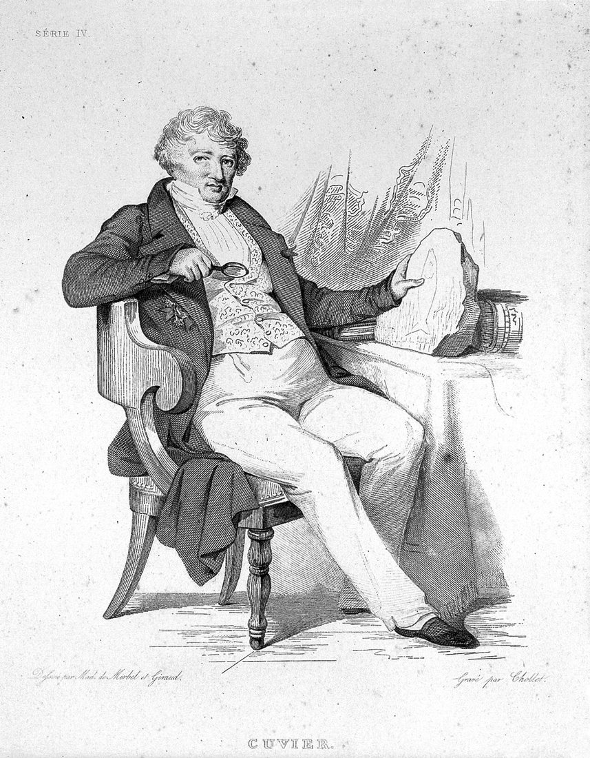
Georges Cuvier was one of the most famous scientists in France. He compared the skeletons and muscles of hundreds of animals, living and fossil, and he proved that whole species had died out. His lectures on comparative anatomy were printed in Paris from 1800. A second edition came out in 1835, after his death, and that is the edition most modern papers cite.
In the lesson on standing, Cuvier asked a simple question. How does a human stay upright on two legs, when every other mammal uses four? He went through the body joint by joint. At the ankle, he said, the weight of the body wants to tip the leg forward, so the calf muscles must hold it back. That is why only humans have real calves.
Then he came to the hip. Here the weight of the trunk wants to fold the body forward over the thighs. So the muscles that pull the hip straight must be strong. And one of them most of all.
"That is why the extensors of the hip, and above all the gluteus maximus, are so large in man, who is the only animal with true buttocks, just as he is the only one with true calves."
« C'est pour cela que ses extenseurs, et surtout le grand fessier, sont si considérables dans l'homme, qui est le seul animal qui ait de véritables fesses, comme il est le seul qui ait de vrais mollets. »
Georges Cuvier, Lessons in Comparative Anatomy, Lesson 7. Our translation.
It was a beautiful argument. Humans stand upright. Humans have the biggest glutes. So the glutes must be what holds us upright. It sounded so obvious that almost nobody checked it.
The problem is that Cuvier never watched the muscle work. Nobody could, yet. He was reasoning from shape, the same way you might guess what a tool does from how it looks. The guess turned out to be wrong, and it took a doctor with a box of batteries to show it.
Part 2 · The test
The doctor with the electrodes
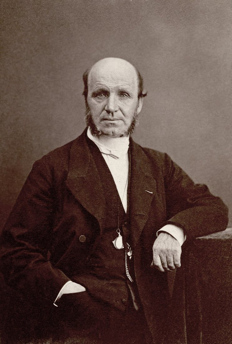
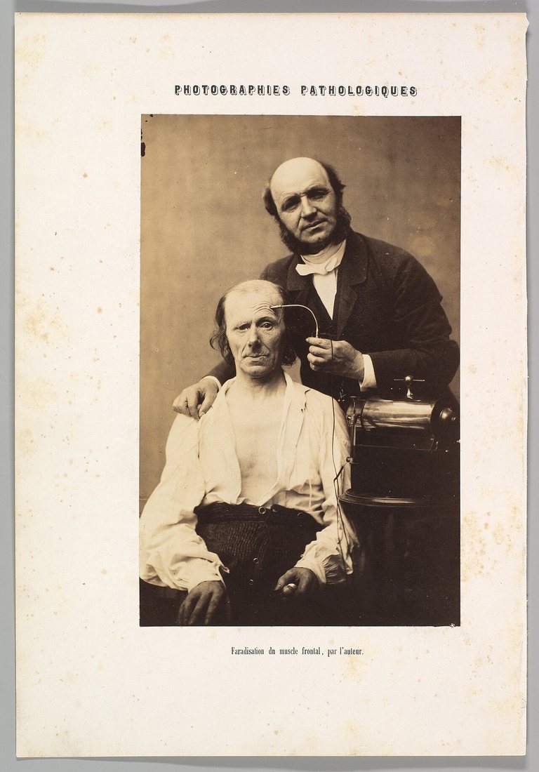
Guillaume Duchenne came from Boulogne, a fishing town on the north coast of France. When he moved to Paris, he added "de Boulogne" to his name, so people would not confuse him with another doctor there. He spent most of his career walking from hospital to hospital, carrying an electrical machine.
His idea was clever. Put two wet electrodes on the skin, right over one muscle, and send a small current through. That one muscle contracts, and nothing else does. Now you can see exactly what it does on its own. He did this for the face first. His 1862 book of photographs showed people smiling, frowning and crying, all made by electricity. Today the real, eye-crinkling smile is still called the Duchenne smile.
Then he turned to the rest of the body. He had a second, even better tool: patients. In the wards of Paris there were people who had lost the use of one muscle, through nerve injury or disease. Each one was a natural experiment. If a person has no working glute max, what can they still do?
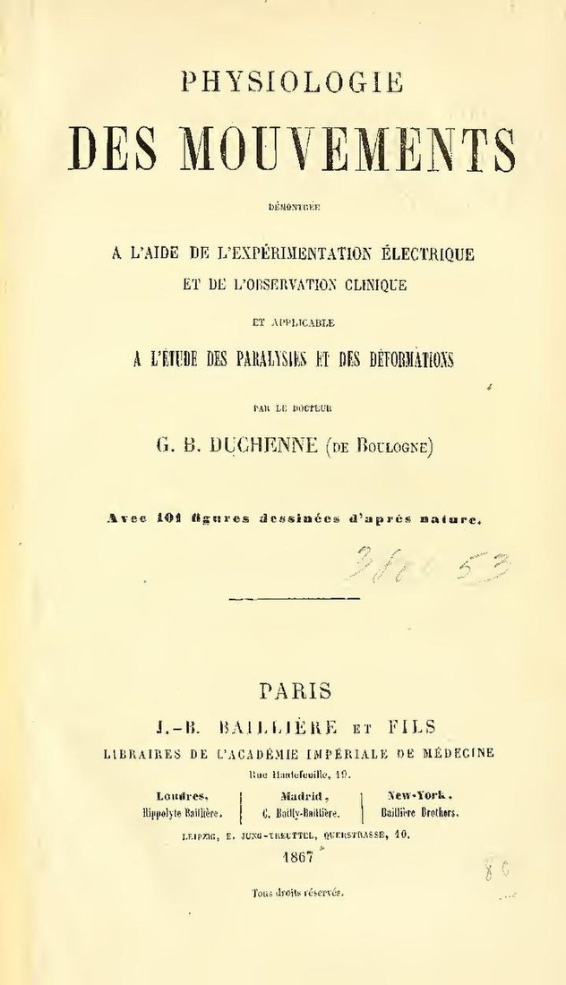
The answer, published in 1867, surprised him. When he sent current into the glute max, it pulled the thigh back very hard and turned it outward. A powerful muscle. But when he watched patients whose glute max was paralysed, they stood normally. They walked on flat ground normally. In quiet standing and in ordinary walking, he concluded, the muscle was doing nothing at all.
So where did they struggle? Duchenne made a list.
Without a working glute max, his patients could still
- Stand
- Walk on flat ground
But they struggled to
- Climb stairs
- Walk uphill
- Rise from a seat
- Jump
- Dance
- Carry a heavy load on the shoulders
Duchenne's list, from Physiology of Movement, 1867. Drawn by Booty Center.
Read that list again. Every item is a big effort: lifting the body up a step or a slope, driving it off the ground, or bracing under a load. Standing still was not on it. For holding the body upright, Duchenne gave more credit to the hamstrings at the back of the thigh than to the glute max.
In other words, Cuvier's idea had already failed its first real test. The biggest muscle in the hip was not a posture muscle. It was a power muscle, saved for the hard jobs.
Where the muscles sit
The glute max is the big outer muscle, highlighted in red. It runs from the back of the pelvis and the base of the spine down to the back of the thigh bone. Under it and to the side sit two smaller glutes, the medius and the minimus. Those two are the stars of the next part of the story.
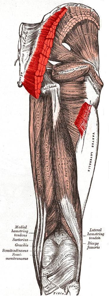
Duchenne also mapped those side glutes. The gluteus medius, he found, works in two halves. The front half turns the thigh inward. The back half turns it outward. And he saw what happens when it is weak: stand the patient on that leg, and the pelvis drops on the other side. He described it in 1867. A surgeon in Germany would make it famous almost thirty years later.
Same man, other fame
A muscle disease that mostly affects boys, Duchenne muscular dystrophy, is named after him. He described it in the 1860s, in the same years he was studying the glutes.
Part 3 · The sign
The waddle and the one-leg test
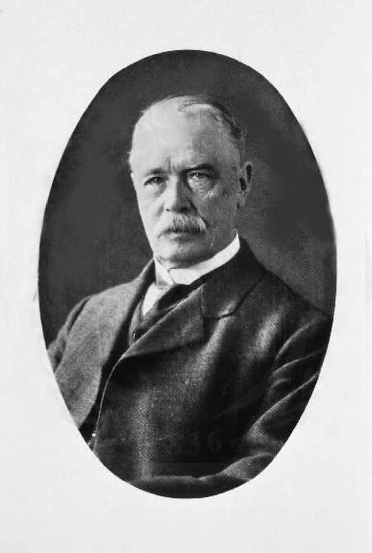
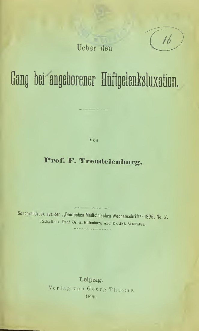
Friedrich Trendelenburg was a surgeon, and a very good one. Surgeons still tilt patients head-down on the operating table in the Trendelenburg position. But in 1895, at the surgical clinic in Bonn, he was puzzling over a walk.
Some children are born with a hip joint that is out of place. These children walked with a rolling waddle, swaying from side to side. Many doctors put the waddle down to the loose joint itself, the thigh bone sliding up and down. Trendelenburg looked closer. He traced it to the side glutes of the standing leg, the medius and the minimus. With the hip out of place, they could not do their job, which is to hold the pelvis level when you stand on one leg.
Think about walking. With every step, for a moment, all your weight is on one leg. In that moment, the side glutes of that leg hold the pelvis from the side and stop the other side from dropping. If they cannot, the pelvis tips, and the body sways to make up for it. Step after step, that becomes a waddle.
His check was so simple that anyone can do it. Ask the person to stand on one leg, and watch the pelvis from behind. Level is good. Dropping on the lifted side means the side glutes of the standing leg are not coping. Clinics around the world still use it, and it still carries his name: the Trendelenburg test.
His paper was written in German. An English translation was published in 1998. You can find both in the sources below.
Part 4 · The scorecard
A century in six dates
- 1773A colour print shows the muscles of the back, after the anatomist Albinus.
- 1800Cuvier's lectures on comparative anatomy are first printed in Paris.
- 1835A new edition appears after his death. Modern papers cite this one.Idea
- 1862Duchenne publishes his photographs of faces moved by electric current.
- 1867Duchenne's Physiologie des mouvements. The glute max rests in standing and walking.Test
- 1895Trendelenburg explains the waddle. The one-leg test is born.Sign
So by 1895 there were three answers on the table. An idea, that the big glute holds us upright. A test, showing it rests during standing and walking and works hard when the body has to climb, rise or jump. And a clinic sign, showing that weak side glutes let the pelvis drop.
Only one of these was a guess. The other two came from watching living people. That difference is the whole story of glute science in one line, and you will see it again in every chapter of this magazine.
The old belief this chapter tests
"The big glute is there to hold us upright."
Not supportedDuchenne's patients stood and walked on flat ground without it in 1867. Yet the upright idea stayed in textbooks for about another hundred years, until scientists could record the muscle directly. That is Chapter 2.
What this means for training today
Your glute max does little while you stand or stroll. It works hardest when your hips must drive the body up or forward against resistance: climbing, rising, jumping, lifting. That is why glute training is built on hip extension against real load. And your side glutes are what keep the pelvis level on one leg, a job they do in every step you take. Later chapters show the modern evidence for both.


