
Chapter 1 ended with a problem. Duchenne could make a muscle twitch with current, and he could watch what patients did without it. But nobody could watch a healthy glute at work, from the inside, while a person stood, walked or climbed. That needed a way to listen to the muscle itself.
The tool that finally did it is called electromyography, or EMG. This chapter tells how it was born, and what happened when scientists in the 1950s to 1980s pointed it at the hip. The short version: the glute was quieter than anyone expected, and it was not one single block.
Part 1 · The spark
Frogs, metal and a twitching arm
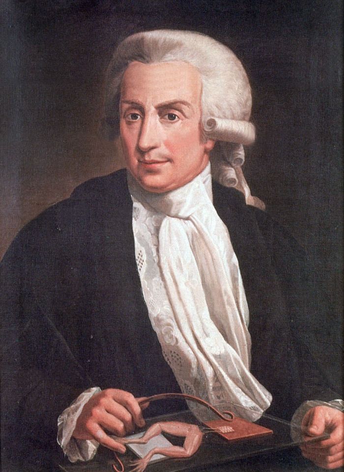
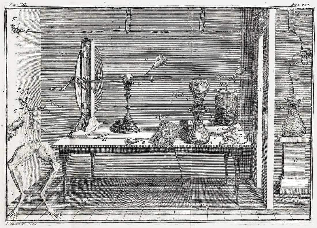
The story starts with frogs. In 1791 the Italian anatomist Luigi Galvani published a book about something strange he saw in his lab in Bologna. Frog legs, prepared for dissection, kicked when they were touched with metal or when an electrical machine sparked nearby. Galvani was convinced that the animal itself made electricity, and that this electricity was what moved the muscles.
He was not fully right about how it worked, and the argument that followed with the physicist Alessandro Volta led to the first battery. But the big idea held: muscles and nerves run on electricity.
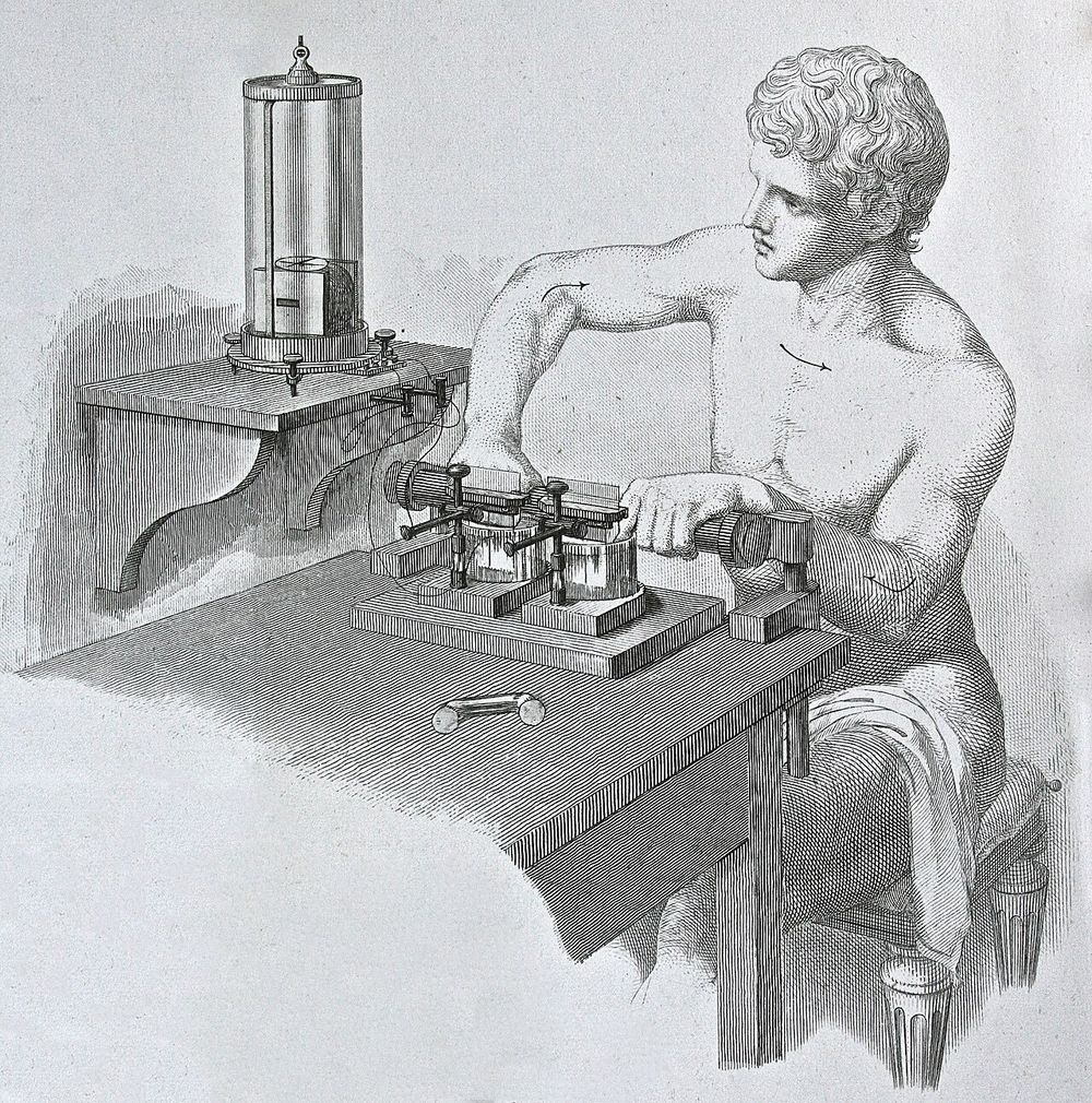
The next step came in Berlin in the 1840s. The physiologist Emil du Bois-Reymond sat at a table, dipped his fingers into two small vessels wired to a very sensitive meter, and tightened the muscles of one arm. The needle moved. A living person's working muscle was giving off a current you could measure.
It was a beautiful proof, but it was crude. A needle swinging on a dial cannot tell you which muscle is working, or how hard, or when. That took another century of better amplifiers, screens and electrodes. By the 1950s, labs could finally record a single muscle in a moving person.
How EMG works, in one line
Every time a muscle fibre contracts, it gives off a tiny electrical signal. An electrode on the skin, or a fine wire placed in the muscle, picks it up. A quiet line means the muscle is resting. A busy line means it is working.
Part 2 · The surprise
The quiet giant
One of the first EMG studies of the hip and thigh muscles in healthy people came out in 1951, from Wheatley and Jahnke. The glutes were now on the record. But the study that really changed the story came six years later.
In 1957, Joseph and Williams placed electrodes on the glute max and asked people to simply stand at ease. If Cuvier was right, and the big glute is what holds us upright, the line should have been busy.
It was almost flat. In relaxed standing, the glute max barely fired at all. They concluded it was not an important posture muscle. It showed moderate activity when people bent forward. Other studies found the same quiet line in ordinary level walking.
Think about what that means. One of the biggest muscles in the human body was mostly switched off during the two things we do most: standing and walking. Duchenne had already hinted at this in 1867 by watching his patients. Now the wires showed it directly.
Part 3 · Mapping the muscle
What makes the glute max fire?
If the glute max is not for standing, what is it for? Through the 1960s, researchers started testing movement after movement to find out.
In 1965, Karlsson and Jonsson mapped what the muscle does. It worked when the hip extended, when the thigh turned outward, and when the leg was lifted out to the side. Later papers cite their study as the first to link the upper part of the glute max to that sideways lift.
In 1968, Fischer and Houtz asked a sharper question: what fires the glute max hardest? Their list, as a later review reports it, was squeezing the muscle hard, turning the thigh outward, lifting the leg sideways against resistance, and driving the thigh and trunk hard into extension while standing.
What fired the glute max hardest, Fischer and Houtz, 1968
- Squeezing the muscle hard
- Turning the thigh outward
- Lifting the leg sideways against resistance
- Hard extension of the thigh and trunk, standing
Notice the pattern. Again, every item on the list is effort. Nothing on it is quiet standing.
Part 4 · Not one block
Upper, lower, front, middle, back
The next discovery changed how we talk about glutes to this day. Bit by bit, the wires showed that these muscles are not single blocks. Different parts of the same muscle can do different jobs.
In 1978, Gary Soderberg and Dostal turned to the side glute, the gluteus medius. They placed a fine wire electrode in the front, the middle and the back of the muscle in ten people. In some tasks, the three parts fired at different times. One muscle, three working parts.
In 1983, a team in California led by Lyons, with the famous gait researcher Jacquelin Perry, recorded eleven healthy people walking and climbing stairs. They found the upper glute max behaved more like the side glute, helping to steady the hip. The lower glute max was the main hip extensor going up stairs. The glute max was not one uniform block.
When a coach today talks about training the "upper glutes" or the "lower glutes", this is the line of research behind the idea, even if the coach has never heard of it.
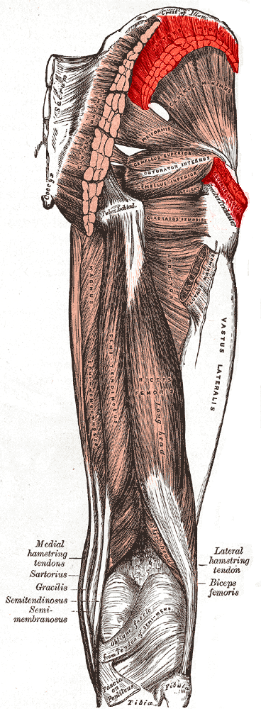
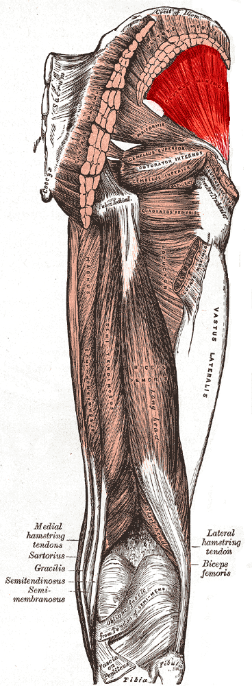
In 1989, Gottschalk, Kourosh and Leveau went one step further and redrew the whole side of the hip as one system. The tensor fasciae latae, a small muscle at the front of the hip, balances the body's weight in walking. The gluteus medius, with its three parts, steadies the hip early in each step. The gluteus minimus steadies it later. Three muscles, one team, each with its moment.
Part 5 · The scorecard
Forty years in seven dates
- 1791Galvani: muscles and nerves run on electricity.
- 1840sDu Bois-Reymond measures a working human muscle.
- 1957Joseph and Williams: the glute max is quiet in standing.Key
- 1965Karlsson and Jonsson map its jobs.
- 1978Soderberg and Dostal: three parts in the medius.
- 1983Lyons and Perry: upper and lower glute max differ.Key
- 1989Gottschalk: the side of the hip as one system.
The old belief this chapter tests
"The glutes hold us upright, and the glute max is one muscle doing one job."
Not supportedThe wires showed a glute max at rest in standing and quiet in level walking. And its upper and lower parts, like the three parts of the side glute, behave differently.
What this means for training today
This is why good glute programs do not rely on standing or walking to train the glute max. It needs effort: climbing, rising, extending the hip against load, and turning or lifting the thigh against resistance. And it is why programs include both hip extension work and side work, because different parts of the glutes respond to different jobs.
But the wires left one big question open. If the glute max rests when we stand and walk, why is the human glute so big? That is Chapter 3.


