What Actually Decides the Shape of Your Butt
MRI and ultrasound studies show where butt fat actually sits, and why bone and muscle set your shape more than fat does.
Almost every woman who trains her glutes has had the same thought. If I get leaner and build more muscle, I will end up with a rounder, fuller shape. It sounds obvious. It is also, in an important way, wrong.
Not wrong because training does not work. It does. Wrong because of what people assume is doing the work. The belief is that roundness comes from the muscle growing and the fat coming off. The imaging evidence points somewhere else, and once you see it, a lot of frustration starts to make sense.
This article walks through what MRI, ultrasound and cadaver anatomy studies actually found about the gluteal region. Where the fat sits. How much of it there is. What changes it. And what the single best study on butt shape discovered when it compared women with aesthetically ideal buttocks against everyone else.
The answer to that last one surprised the surgeons who ran it.
Start with the four layers
Everything you see from the outside is four things stacked on top of each other. From the inside out: the pelvis bone, the gluteal muscles, a layer of fat under the skin, and the skin itself.
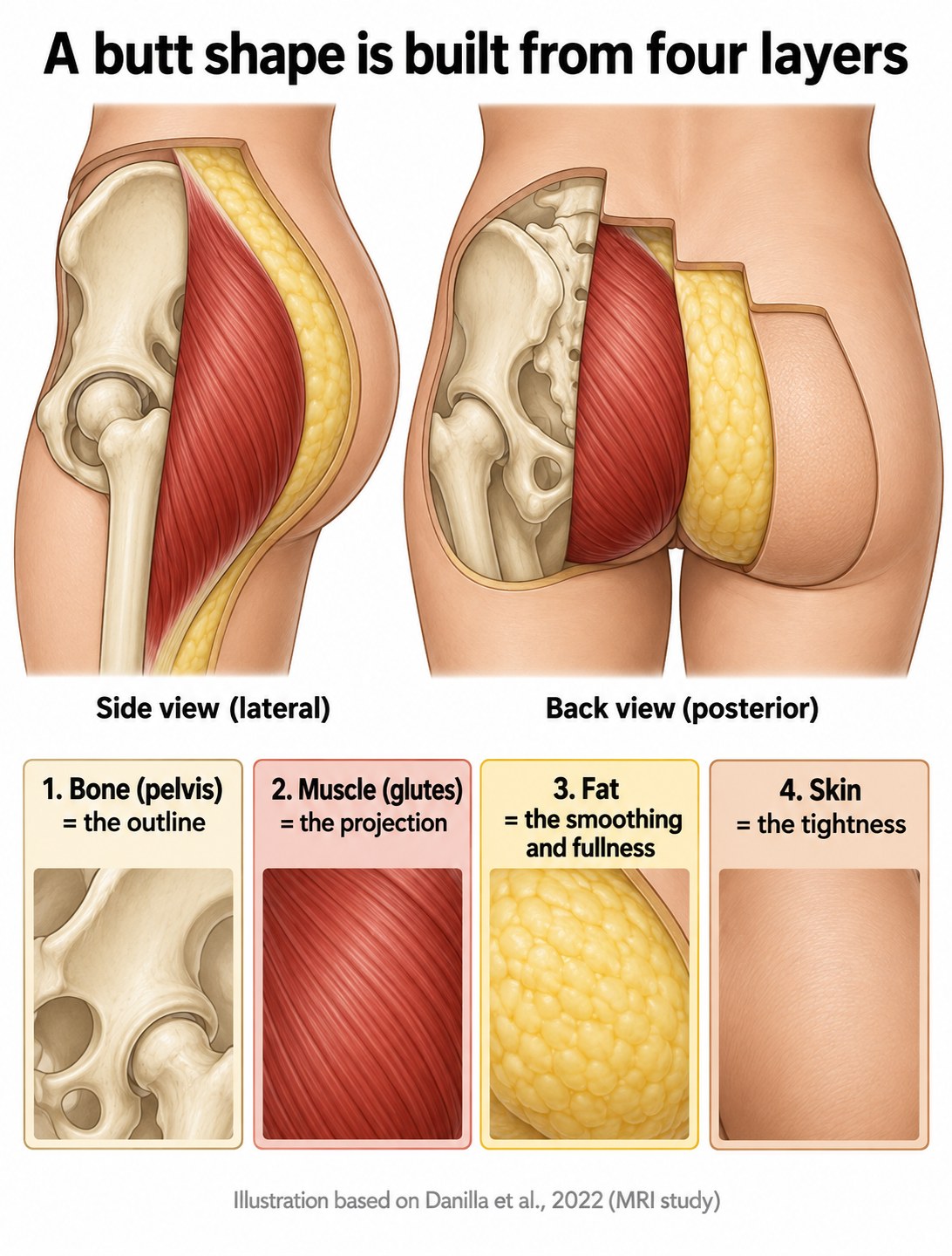 The four layers that build a butt shape, in side view and back view.
Each one does a different job. The bone sets the outline and the width. The muscle sets how far the shape projects backward. The fat smooths and fills. The skin holds it all and decides how tightly it drapes.
Keep those four jobs in mind, because the rest of this article is really about which of them you can change and which you cannot.
Where the fat actually sits
Most people picture gluteal fat as an even blanket, thicker on some women and thinner on others, but spread uniformly. It is not.
A 2022 anatomy study in Clinical Anatomy measured subcutaneous fat thickness across the buttocks in 30 cadavers. The fat was thickest in the centre of each buttock and thinnest out on the lateral side, toward the hip. That is measured, not estimated.
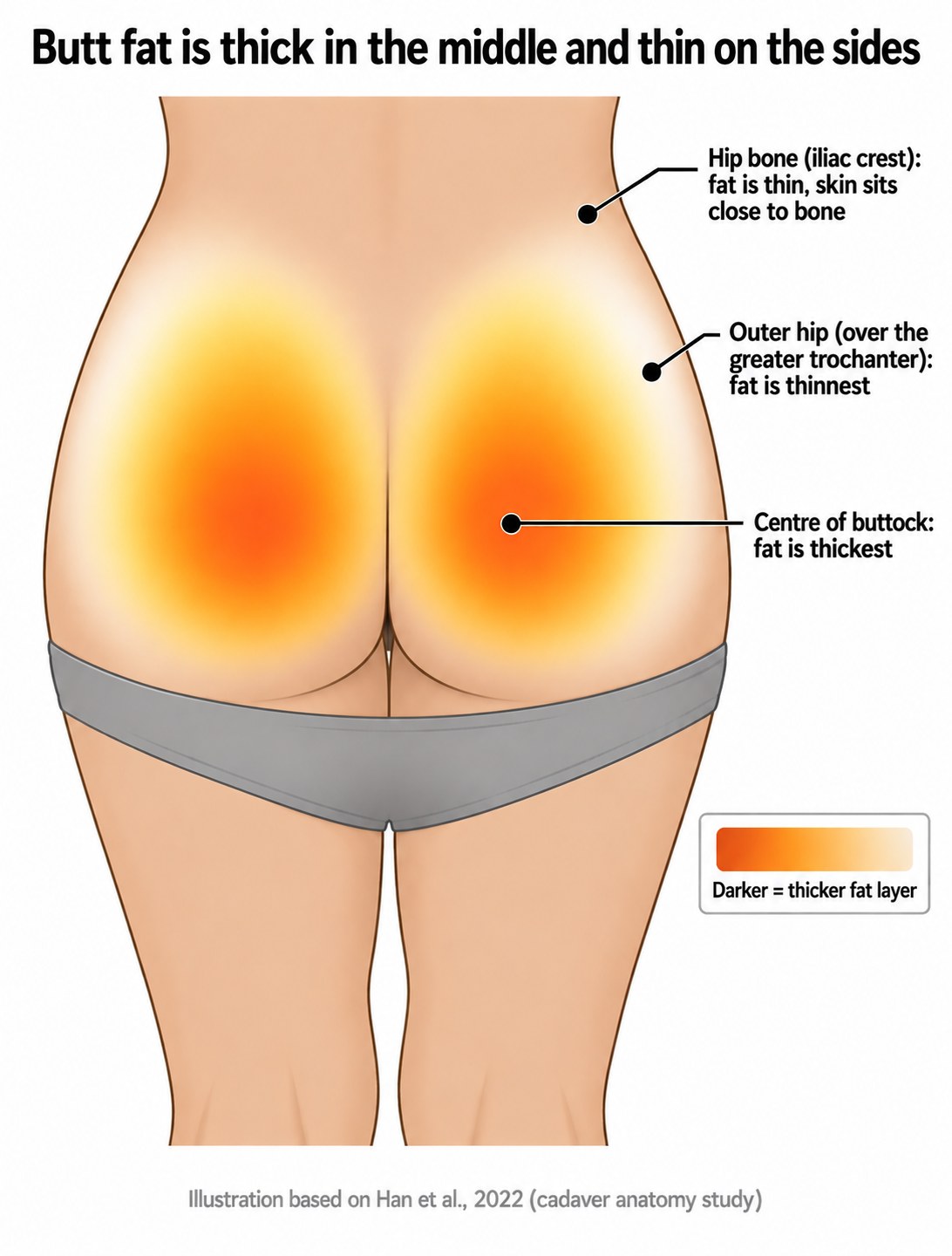 Fat thickness across the buttock, based on Han and colleagues, 2022.
This one finding explains more than it looks like it should. The outer hip, the area where people notice hip dips, has very little fat over it to begin with. The bone and the smaller gluteus medius muscle sit close to the surface there. Meanwhile the centre of the cheek carries the deepest cover.
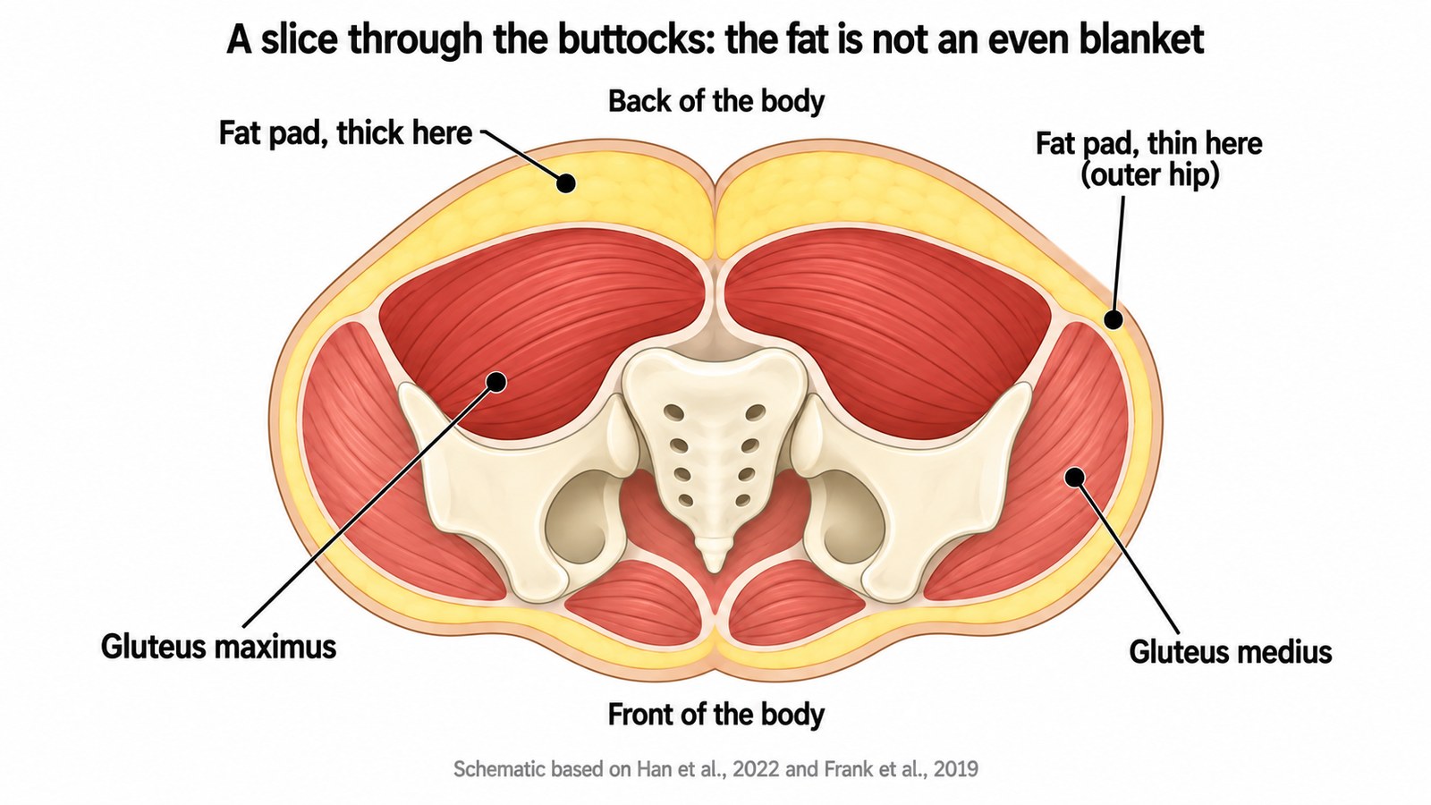 A horizontal slice through the buttocks showing the uneven fat layer.
Look at the slice. The pale yellow band is thick at the back and thins out toward the sides. That shape is not something you chose and it is not something training changes.
The fat itself has layers
A 2024 study used 7 Tesla MRI, a much higher resolution than a standard clinical scanner, along with tissue microscopy, to look at the gluteal and thigh fat in detail. It found three distinct layers rather than one mass: a superficial layer of small upright fat lobules held by vertical fibrous bands, a middle layer, and a deep layer of larger, flatter lobules.
The same study described small fat structures called adipose papillae that rise from the skin layer down into the superficial fat. These are part of why the surface of the skin looks the way it does.
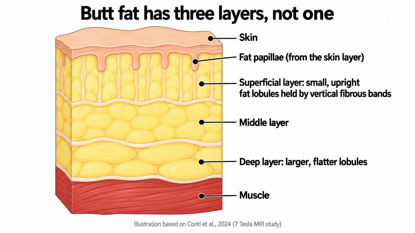 The three layers inside gluteal fat, based on Conti and colleagues, 2024.
Why this matters: the layers do not behave the same way, which is the next point.
What controls how much fat is there
A 2019 study in Plastic and Reconstructive Surgery measured gluteal fat with ultrasound in 150 adults, evenly split between men and women, across five age decades and three body mass index groups.
The main finding is simple. In women, every 1 point of body mass index corresponded to about 4 more millimetres of total gluteal subcutaneous fat. In men it was about 3 millimetres.
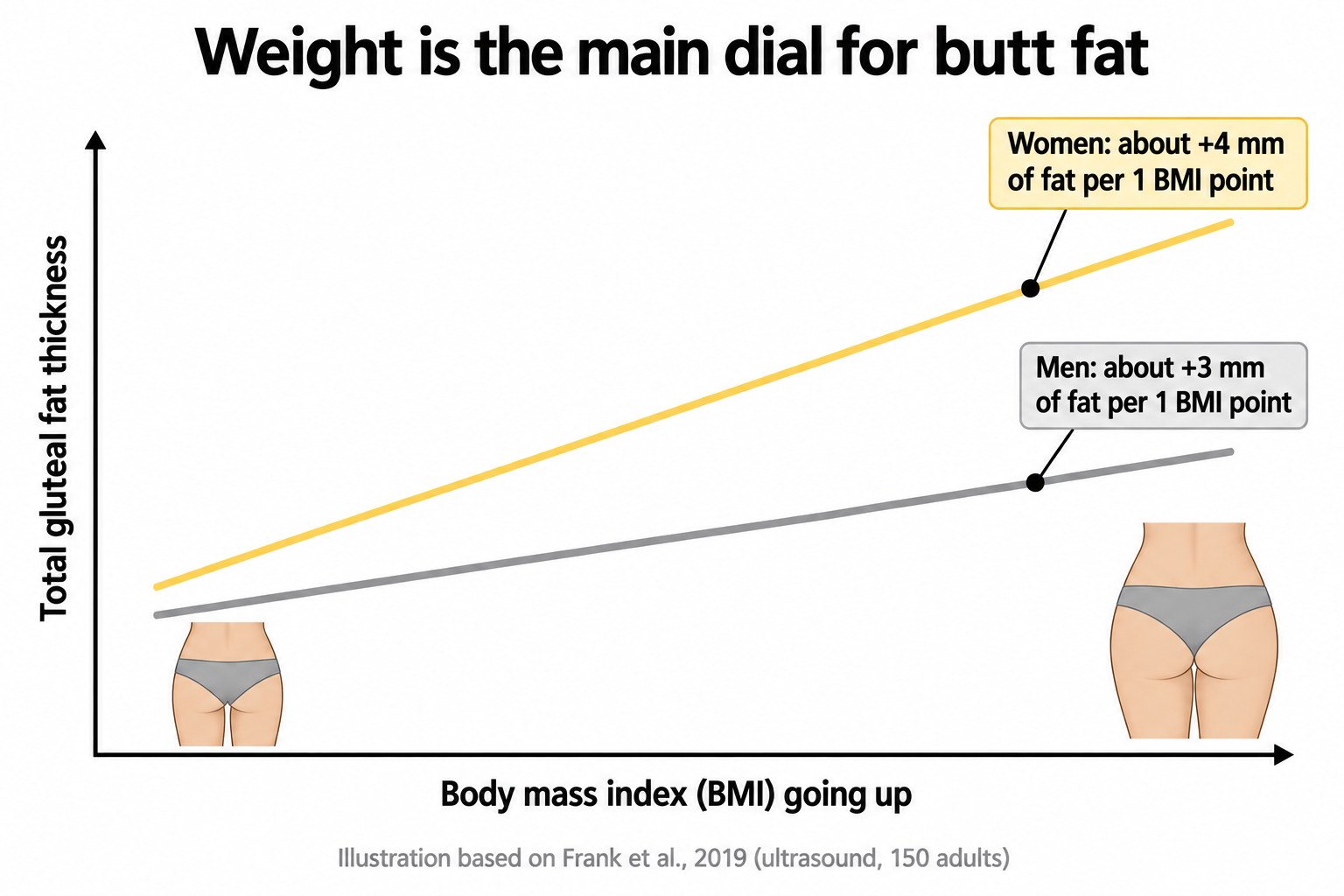 Gluteal fat thickness rises with body weight, based on Frank and colleagues, 2019.
A separate MRI study of 133 women aged 15 to 80 found the same relationship. Body mass index was moderately to strongly correlated with total gluteal fat thickness, and age on its own showed no correlation with it.
So body weight is the main dial. Not age, not genetics in the loose way people use that word, and not the type of training you do.
Age and weight do not add fat in the same place
The 2019 ultrasound study found something more specific. As body mass index went up, the superficial layer thickened most. As age went up, the deep layer thickened.
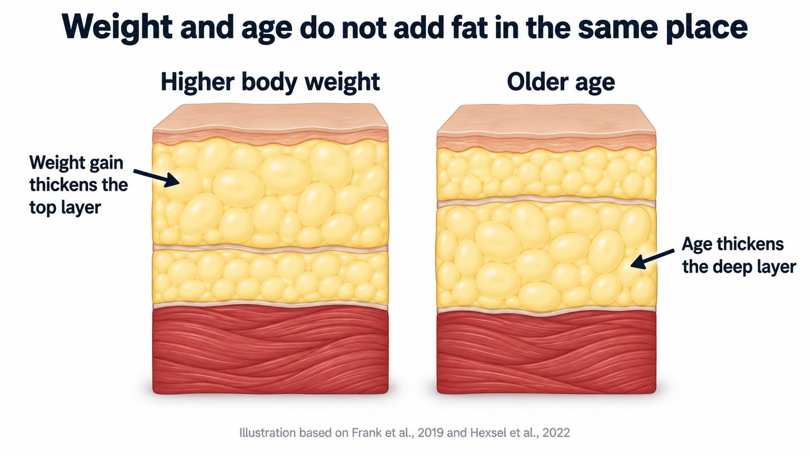 Weight gain thickens the top layer. Age thickens the deep layer.
Same total amount of fat, different position within the stack, different look on the surface. This is one reason two women at the same weight and the same age can look different.
The study that should change the conversation
In 2022, a group published a case control study in Aesthetic Plastic Surgery. They took women who work professionally as buttock models, meaning women whose buttocks are considered aesthetically ideal by the market that pays for them, and compared them against women with normal buttocks. They used MRI, standardised photography and direct body measurements.
Here is what they found. Compared to the control group, the models had a narrower waist, a narrower iliac crest, wider hips, and a bigger and thicker gluteus maximus muscle.
And the amount of subcutaneous fat was equal in both groups.
The fat was the same. The bone and the muscle were different.
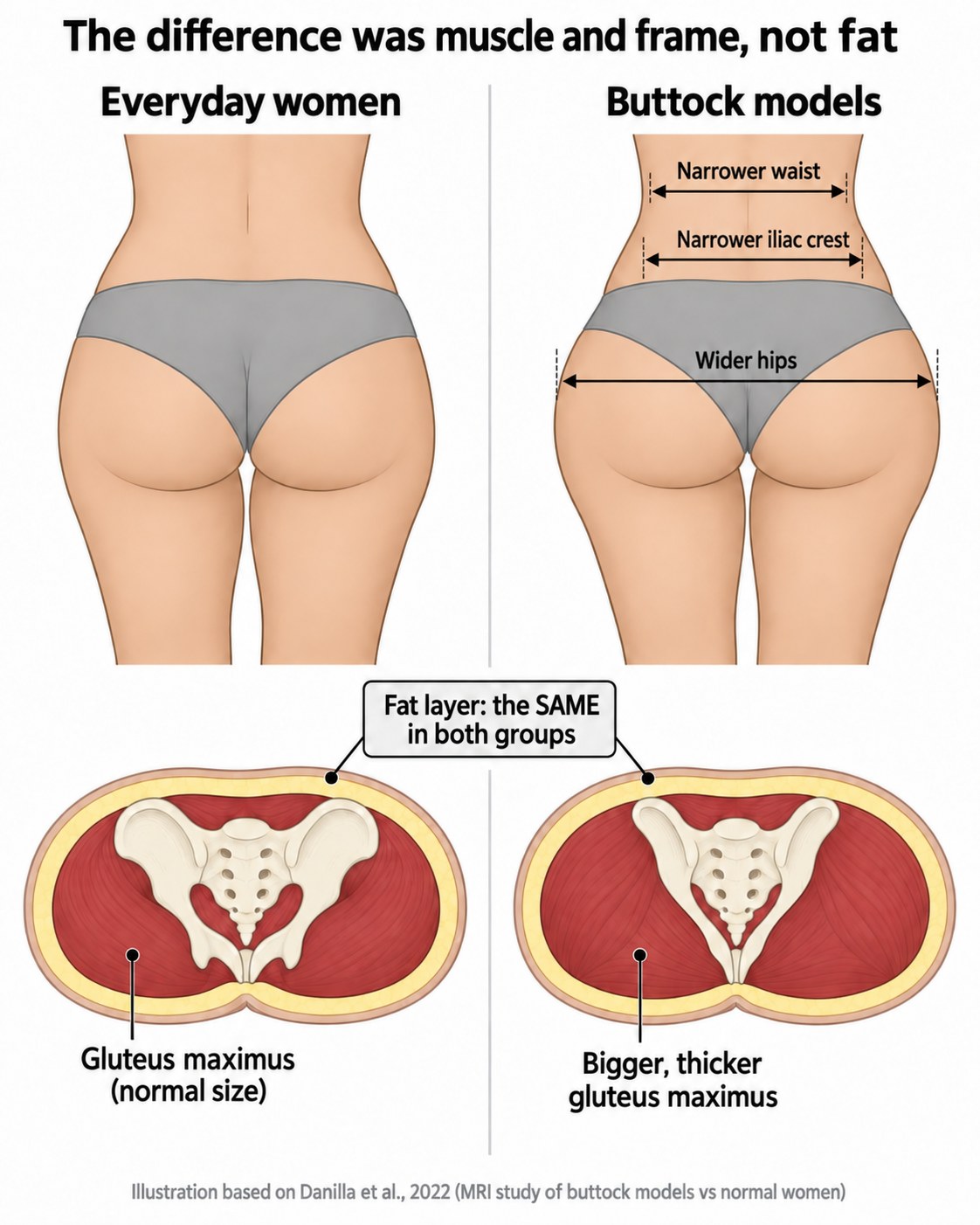 Buttock models compared to everyday women, based on Danilla and colleagues, 2022.
Read that again, because it undercuts the most common assumption in this whole topic. The shape people are chasing was not produced by having more fat in the right places, and it was not produced by having less fat either. It was produced by the skeleton underneath and the size of the muscle sitting on it.
That is the honest version of the answer. It is less marketable than the usual one. It is also more useful, because it tells you where to put your effort and what to stop blaming yourself for.
Your frame is bone, and bone does not diet or train
Plastic surgeons who do gluteal contouring have been classifying the bony frame for years. The most cited system, published in 2006, sorts patients by frame type, muscle type and the relationship between the two.
One point of caution here, and it matters. These frame categories come from surgical practice, not from population measurement studies. They are a useful way to describe what surgeons see, not validated types with data behind them. In fact, a 2022 systematic review searched the plastic surgery literature and concluded that no standardised, reproducible classification system for gluteal size and shape exists at all.
So treat the categories as descriptive language rather than as a diagnosis of what you are.
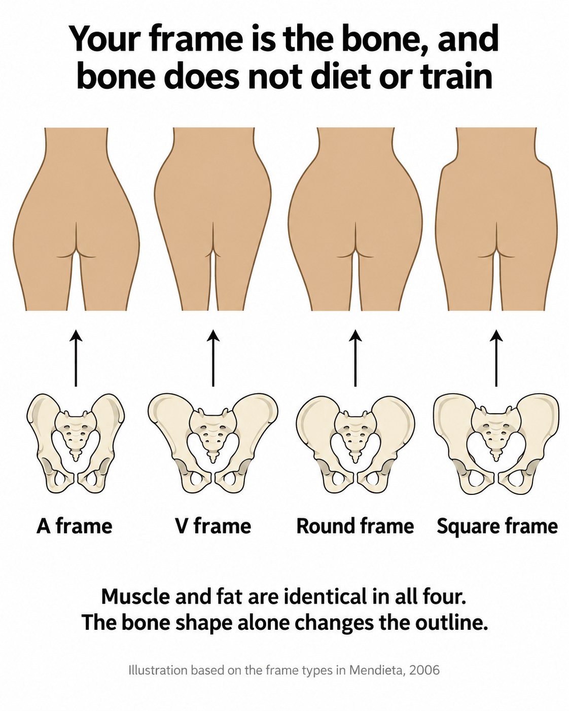 Four frame descriptions used by surgeons, with the pelvis shape behind each outline.
With that caveat in place, the underlying principle holds up and it is anatomically straightforward. The width of your iliac crest, the distance between your hip bones, and the spacing between the crest and the top of your thigh bone set the outline of your hips. Those measurements are fixed in adulthood.
Same fat, same muscle, different outline
Put two women side by side with identical fat thickness and identical muscle size. One has a narrow iliac crest with the widest point lower down. The other has a wide, high crest with straight sides. They will not look the same from behind, and no amount of training will swap one for the other.
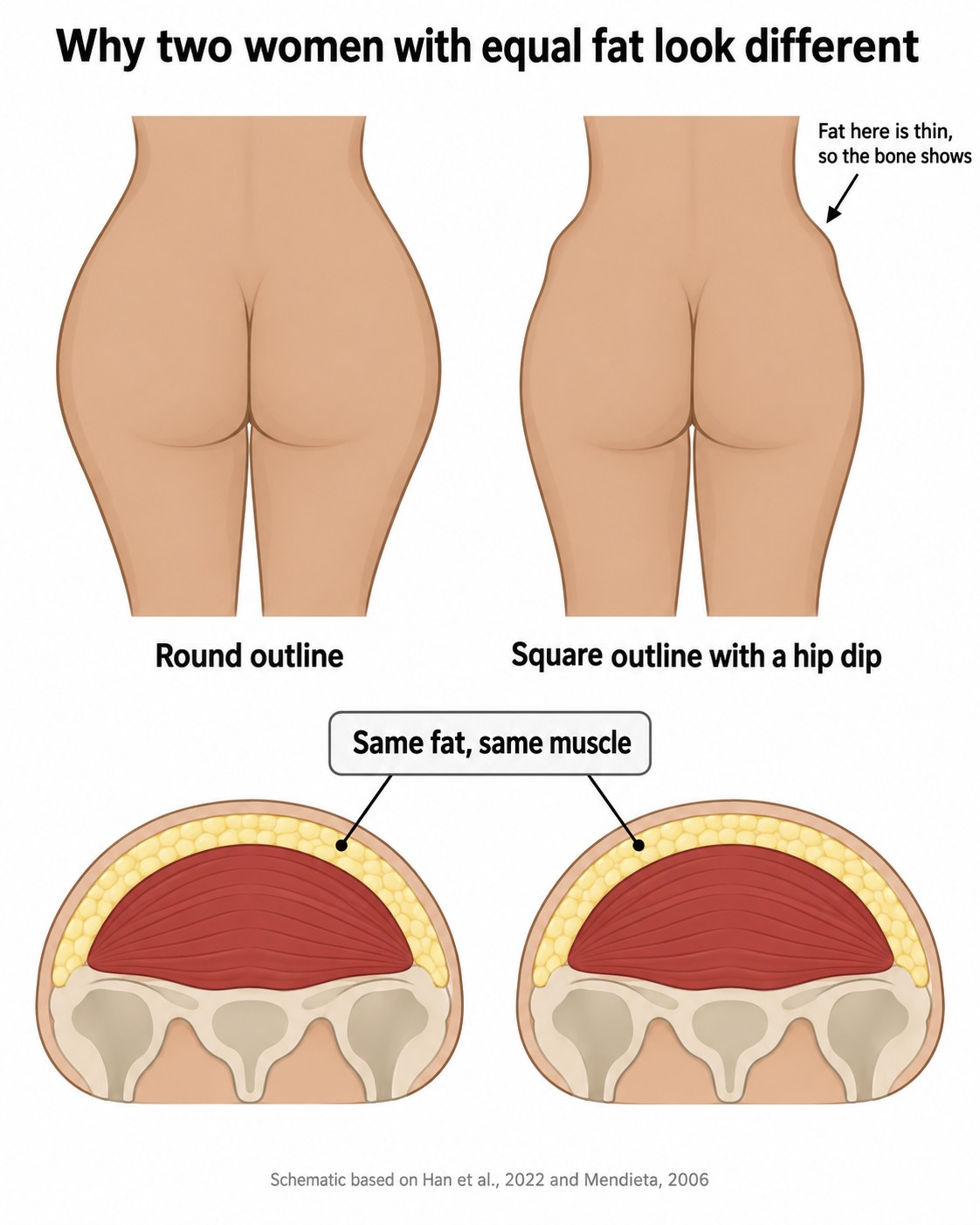 Two outlines with equal fat and equal muscle underneath.
The reason the difference shows so clearly goes back to Figure 2. Fat is thin over the outer hip. There is not enough cover there to soften a wide bony frame or fill a gap between bones.
What really happens when you get leaner
Now the part people most want to know.
As body fat drops, gluteal fat drops with it, because the two move together. The outline gets smaller. But the shape that emerges is the shape of the bone and muscle underneath, and that shape was always there.
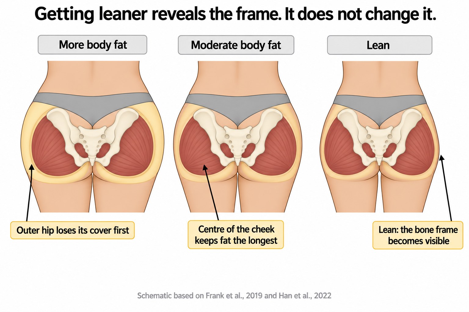 The same hips at three levels of body fat, with the bone and muscle held identical.
Two things happen in a predictable order. The outer hip loses its thin cover first, so any gap between the hip bone and the thigh bone becomes more visible as you lean out. The centre of the cheek holds its fat longest, because that is where the layer is deepest to start with.
This is why some women get leaner and feel like the shape got worse rather than better. Nothing went wrong. The cover came off a frame that was always underneath it.
Getting leaner reveals the frame. It does not change it.
That is not an argument against getting leaner. It is an argument for knowing what leanness will and will not do, so the result does not come as a disappointment.
What muscle growth actually does
The gluteus maximus is the one layer in this stack that responds directly to training, and it is the layer that made the difference in the buttock model study.
What it changes is projection. A bigger gluteus maximus pushes the shape further back in side view and lifts the point of maximum projection higher. In the model group, that peak sat about 2.7 centimetres above the level of the pubic bone.
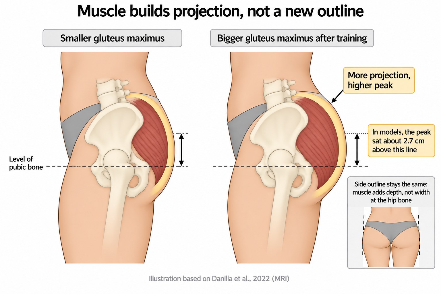 What a larger gluteus maximus changes, and what it does not.
What it does not change is your width at the hip bone or the outline of your frame from behind. Muscle adds depth. It does not add bone.
So the realistic promise of glute training is a fuller, higher, more projected shape on the frame you have. That is a real and worthwhile result. It is just not the same as changing your frame.
The summary, in one table
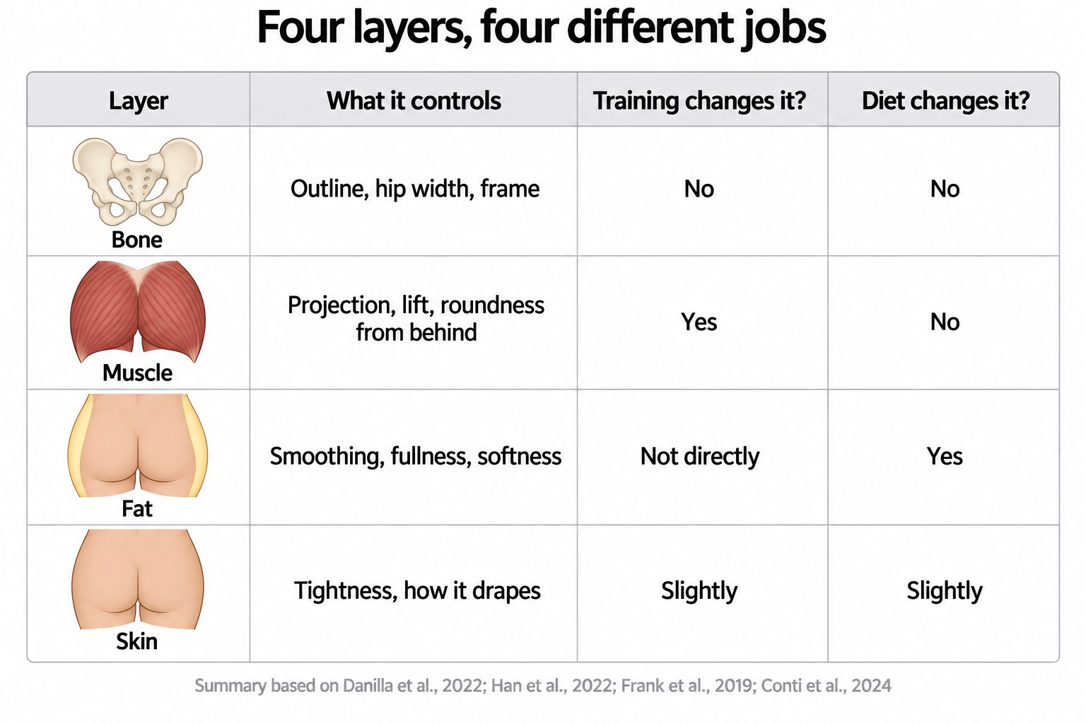 Which layer controls what, and what actually changes it.
If you take one thing from this article, take that table. Most of the confusion in this topic comes from people attributing a result to the wrong layer.
Women, men and why the fat is there at all
A 2023 MRI study compared 45 female and 42 male recreational cyclists, all of whom had ridden more than 7,000 kilometres in the previous year. Matching people this closely for training is unusual and it makes the comparison worth something.
The women had significantly more gluteal subcutaneous fat than the men. They had the same amount of fat inside the muscle. And when glute muscle volume was adjusted for body weight, there was no difference between the sexes at all.
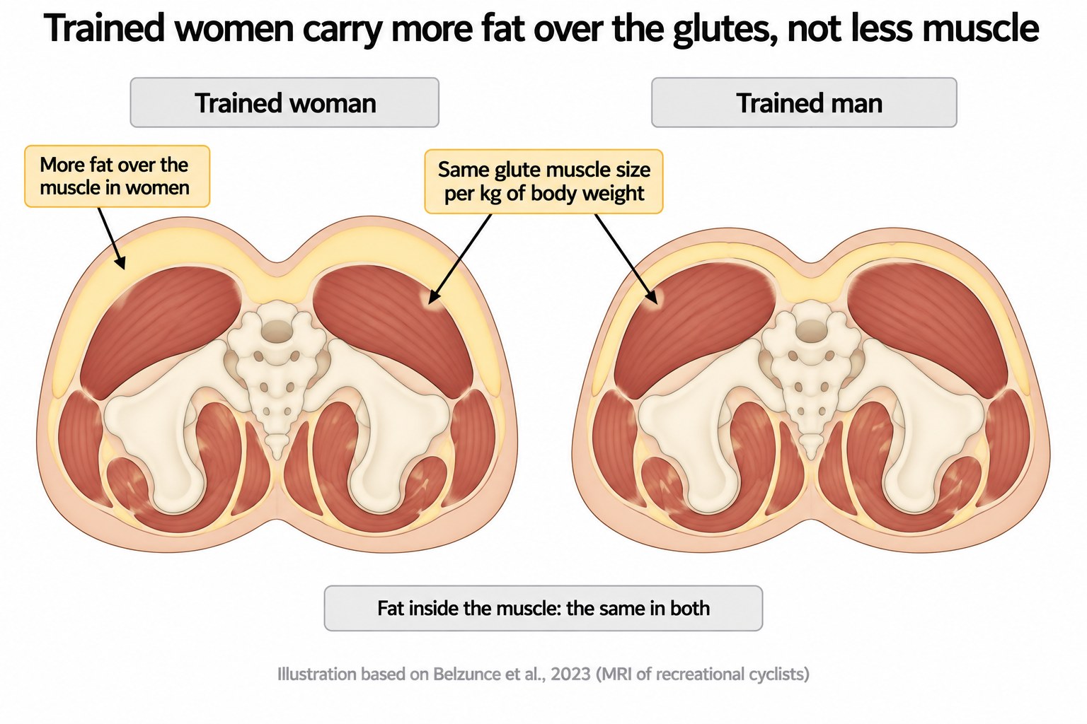 Trained women compared to trained men, based on Belzunce and colleagues, 2023.
So trained women are not carrying less muscle relative to their size. They are carrying more fat over it. That is a normal female pattern, not a training failure.
Why hip and thigh fat resists dieting
Fat depots are not interchangeable. Abdominal fat is metabolically busy, taking fat in and releasing it quickly through the day. Gluteofemoral fat, meaning the fat on the hips and thighs, behaves as a long term store. A widely cited 2010 review in the International Journal of Obesity described it as comparatively passive in day to day metabolism, working instead by long term fatty acid storage.
Estrogen is a major reason storage is directed there in the first place.
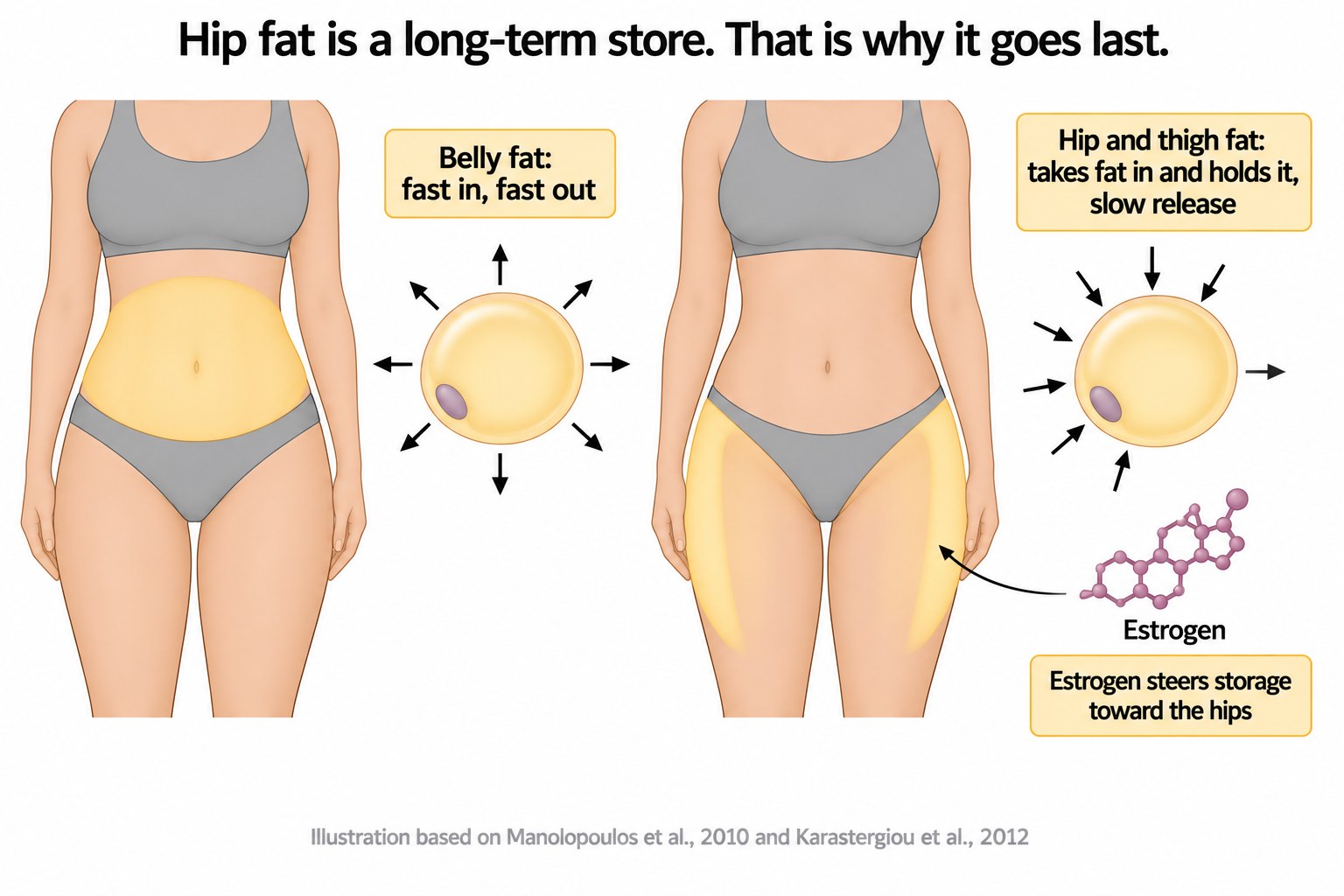 Belly fat and hip fat do not behave the same way.
Practically, this means hip and thigh fat tends to be the last to go in a fat loss phase. Not because anything is broken, but because that depot is built to hold rather than release.
What changes at menopause
When estrogen falls, storage shifts. The pattern moves from gynoid, meaning hip and thigh dominant, toward android, meaning abdominal. This shift is well documented and can be partly reversed with hormone replacement therapy.
A 2002 study in the Journal of Clinical Endocrinology and Metabolism added a detail that fits. In postmenopausal women, basal lipolysis in gluteal fat cells was 77 percent lower than in perimenopausal women matched for body fat. Lipoprotein lipase activity, which drives fat storage, was higher in the gluteal region than the abdominal region in both groups.
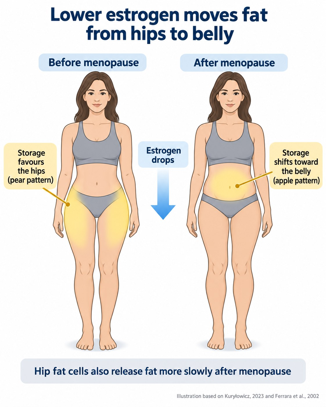 How the storage pattern changes as estrogen falls.
So gluteal fat becomes harder to mobilise after menopause, while new storage trends toward the middle. Worth knowing if you are training through that transition and wondering why the same approach stopped producing the same result.
Hip dips are bone spacing
This deserves its own section because of how much bad advice exists around hip dips.
The dip sits between the top of the hip bone, called the iliac crest, and the greater trochanter, which is the bony bump at the top of your thigh bone. Between those two points sits the gluteus medius, a relatively small muscle, with a thin fat cover over it.
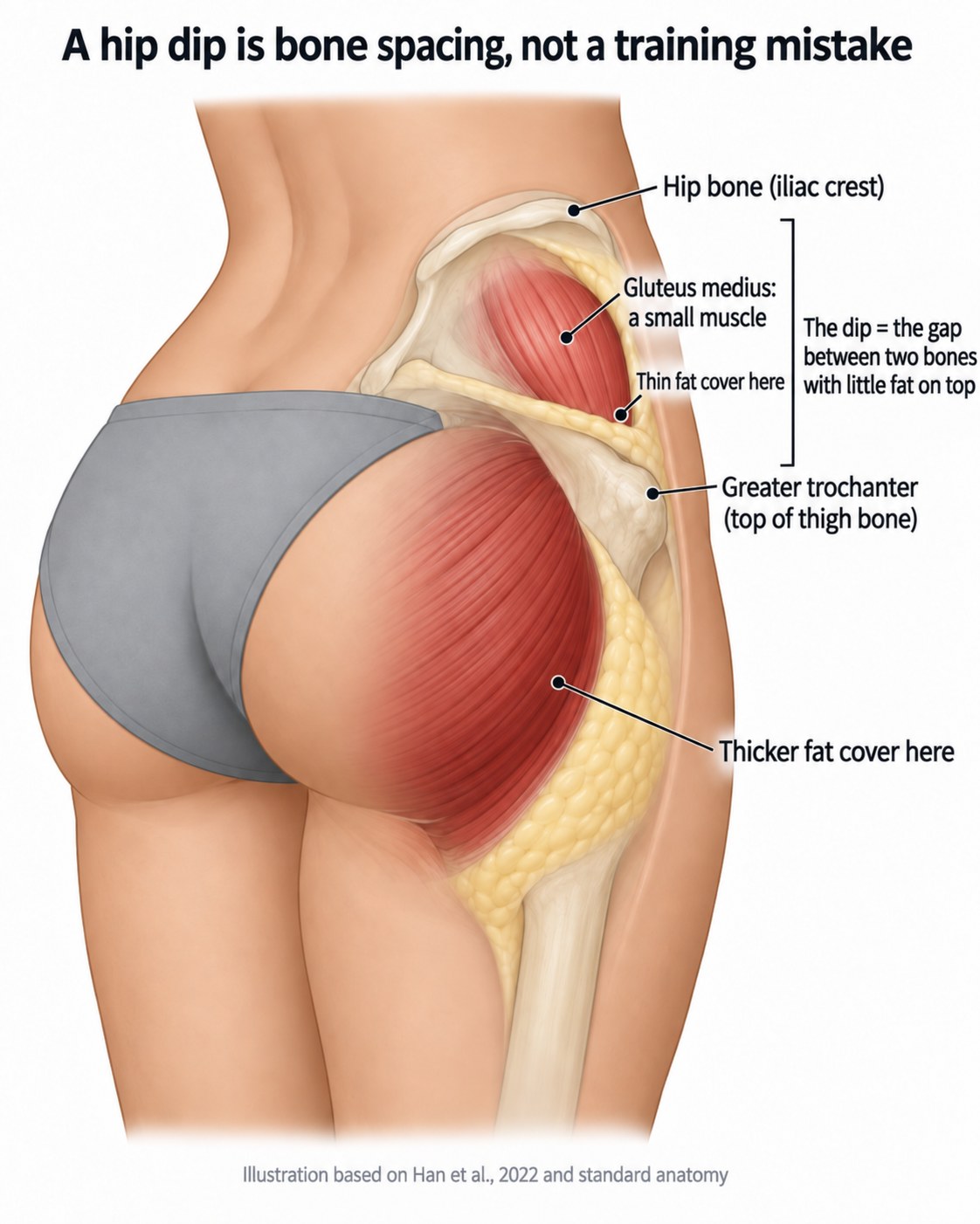 What is underneath a hip dip.
Put those facts together. The distance between two bones is fixed. The muscle in between is small. The fat cover there is the thinnest anywhere on the buttock, as measured in the 2022 cadaver study. A dip in that location is a consequence of bone spacing, not of insufficient training.
Building the gluteus medius can add a small amount of tissue in that window and it is worth training for function regardless. It will not close a gap set by skeletal geometry.
The hidden fibres that hold the shape
The 2022 cadaver study found structures most people have never heard of, and they explain some things that fat and muscle alone cannot.
Superficial fascia, a fibrous sheet within the fat, was present only in the upper buttock and became indistinct lower down. Over the sacrum and tailbone, the skin was bound tightly to the bone as a single mass. And a strong fibre was found running from the sitting bone, the ischial tuberosity, outward into both the gluteus maximus and the skin.
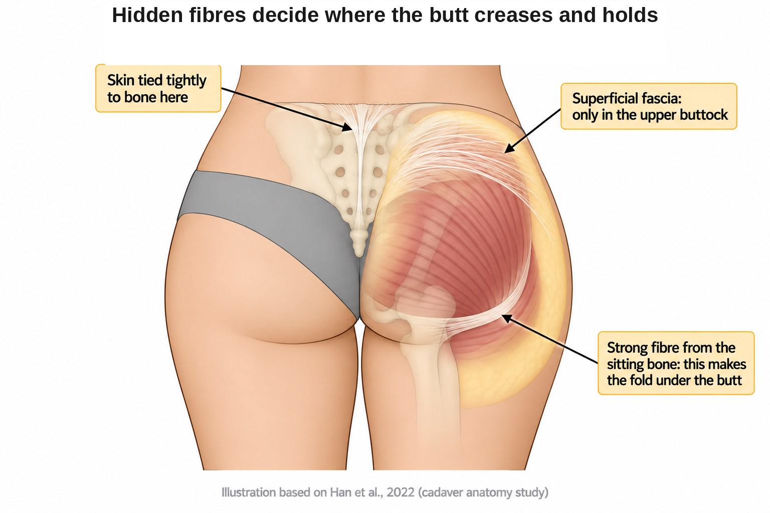 The fibrous structures that decide where the butt creases and holds.
That last fibre is what creates and anchors the fold under the buttock. It is a structural feature, not something produced by muscle tone. Where your fold sits, and how defined it is, is largely decided by these attachments.
What nobody has measured yet
Here is an honest admission that most content on this subject will not give you.
We know fat is thicker in the centre and thinner on the sides, from cadavers. We know total thickness scales with body weight, from ultrasound and MRI. We know models and everyday women carry the same amount of fat, from MRI.
What no study has done is map fat thickness point by point across the buttock in living women, and compare that map between lean trained women and untrained women. That map does not exist in the published literature.
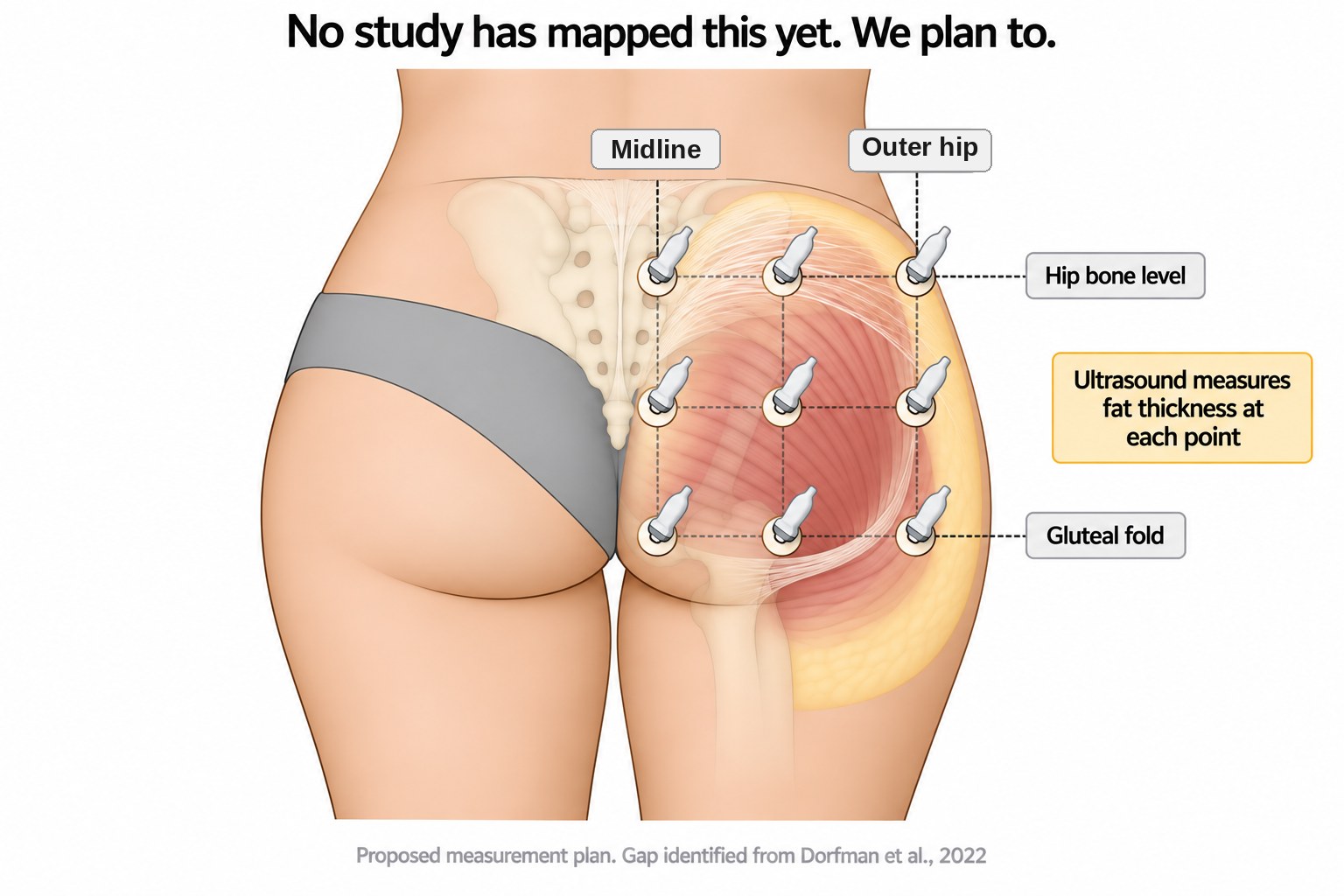 The measurement plan for the map that is missing.
Nine ultrasound measurement points per buttock, running from hip bone level down to the gluteal fold and from the outer hip across to the midline. That is a straightforward study to run and it would answer questions that are currently answered with guesswork.
We intend to run it.
What this means for you
Pulling the whole thing together.
- Your frame sets the outline. Iliac crest width, hip width and the gap between your hip bone and thigh bone decide the silhouette. This is fixed and it is not a flaw.
- Muscle sets the projection. The gluteus maximus is the layer training changes, and in the one study comparing ideal buttocks against normal ones, muscle size was a defining difference.
- Fat smooths and fills, and it scales with body weight. About 4 millimetres of gluteal fat per body mass index point in women.
- Fat is not evenly spread. Thickest in the centre, thinnest over the outer hip. Leaning out reveals the outer hip first.
- Getting leaner does not make a square frame round. It makes the existing frame more visible.
- Hip and thigh fat goes last, by design. That depot is built for long term storage, and it becomes harder to mobilise after menopause.
- Hip dips are bone spacing with thin fat cover. Not a training mistake.
The practical takeaway is not that effort is pointless. It is the opposite. Training the gluteus maximus is the highest leverage thing available to you, because it is the one layer in the stack that responds. Getting leaner is worth doing for plenty of reasons. Just go in knowing that the outline you end up with belongs to your skeleton, and that this was never something you did wrong.
Most of the frustration in this area comes from chasing a result that was assigned at birth while ignoring the one that is genuinely available.
Common questions
Does losing fat make your butt rounder?
No. Losing fat makes the outline smaller, but the shape that emerges belongs to the bone and muscle underneath. An MRI study comparing buttock models to everyday women found both groups had the same amount of subcutaneous fat. The difference was bone and muscle, reported by Danilla and colleagues in Aesthetic Plastic Surgery in 2022.
What causes hip dips?
A hip dip is the gap between the iliac crest and the greater trochanter, with a small gluteus medius and a thin fat cover in between. The distance between those two bones is fixed. The fat over the outer hip is the thinnest anywhere on the buttock, as measured by Han and colleagues in Clinical Anatomy in 2022.
Can you change the shape of your butt?
You can change how far it projects, not the frame it sits on. A larger gluteus maximus pushes the shape further back and lifts the peak, and that is the layer training changes. It does not change your width at the hip bone.
Where is butt fat thickest?
Butt fat is thickest in the centre of each cheek and thinnest over the outer hip. A 2022 anatomy study in Clinical Anatomy measured that pattern in 30 cadavers. The outer hip has very little fat over it to begin with.
Do women have less glute muscle than men?
No. When glute muscle volume was adjusted for body weight, trained women and trained men showed no difference. A 2023 MRI study of recreational cyclists in BMJ Open Sport and Exercise Medicine found the women carried more subcutaneous fat over the muscle, not less muscle for their size.
Why is hip and thigh fat so hard to lose?
Hip and thigh fat is built as a long term store, so it tends to be the last to go in a fat loss phase. Abdominal fat takes fat in and releases it quickly, while gluteofemoral fat is comparatively passive day to day. A 2010 review in the International Journal of Obesity described that pattern.
References
Conti G, et al. Gluteal femoral subcutaneous and dermal adipose tissue in female. Journal of Cosmetic Dermatology. 2024.
Danilla S, et al. What Makes a Beautiful Buttock Beautiful? A Case-Control Study Comparing Buttocks Models versus Normal Women by Magnetic Resonance Imaging, Photography and Anthropometry. Aesthetic Plastic Surgery. 2022.
Dorfman R, et al. Gluteal Shape Framework for Augmentation Surgery: A Systematic Review of the Literature and Proposed Classification System. Plastic and Reconstructive Surgery. 2022.
Belzunce MA, et al. Similarities and differences in skeletal muscle and body composition between sexes: an MRI study of recreational cyclists. BMJ Open Sport and Exercise Medicine. 2023.
Ferrara CM, et al. Differences in adipose tissue metabolism between postmenopausal and perimenopausal women. Journal of Clinical Endocrinology and Metabolism. 2002.
Frank K, et al. Influence of Age, Gender and Body Mass Index on the Thickness of the Gluteal Subcutaneous Fat: Implications for Safe Buttock Augmentation Procedures. Plastic and Reconstructive Surgery. 2019.
Han A, et al. Gross anatomical study of the subcutaneous structures that create the three-dimensional shape of the buttocks. Clinical Anatomy. 2022.
Hexsel D, et al. Gluteal subcutaneous adipose tissue in women of different ages and body mass index observed by magnetic resonance imaging. Journal of Cosmetic Dermatology. 2022.
Karastergiou K, et al. Sex differences in human adipose tissues: the biology of pear shape. Biology of Sex Differences. 2012.
Kurylowicz A. Estrogens in Adipose Tissue Physiology and Obesity-Related Dysfunction. Biomedicines. 2023.
Manolopoulos KN, et al. Gluteofemoral body fat as a determinant of metabolic health. International Journal of Obesity. 2010.
Mendieta CG. Classification system for gluteal evaluation. Clinics in Plastic Surgery. 2006.